
CardaxEP Twin
Build imaging-informed heart models and study how anatomy, scar, and mechanics affect simulated rhythms.
macOS
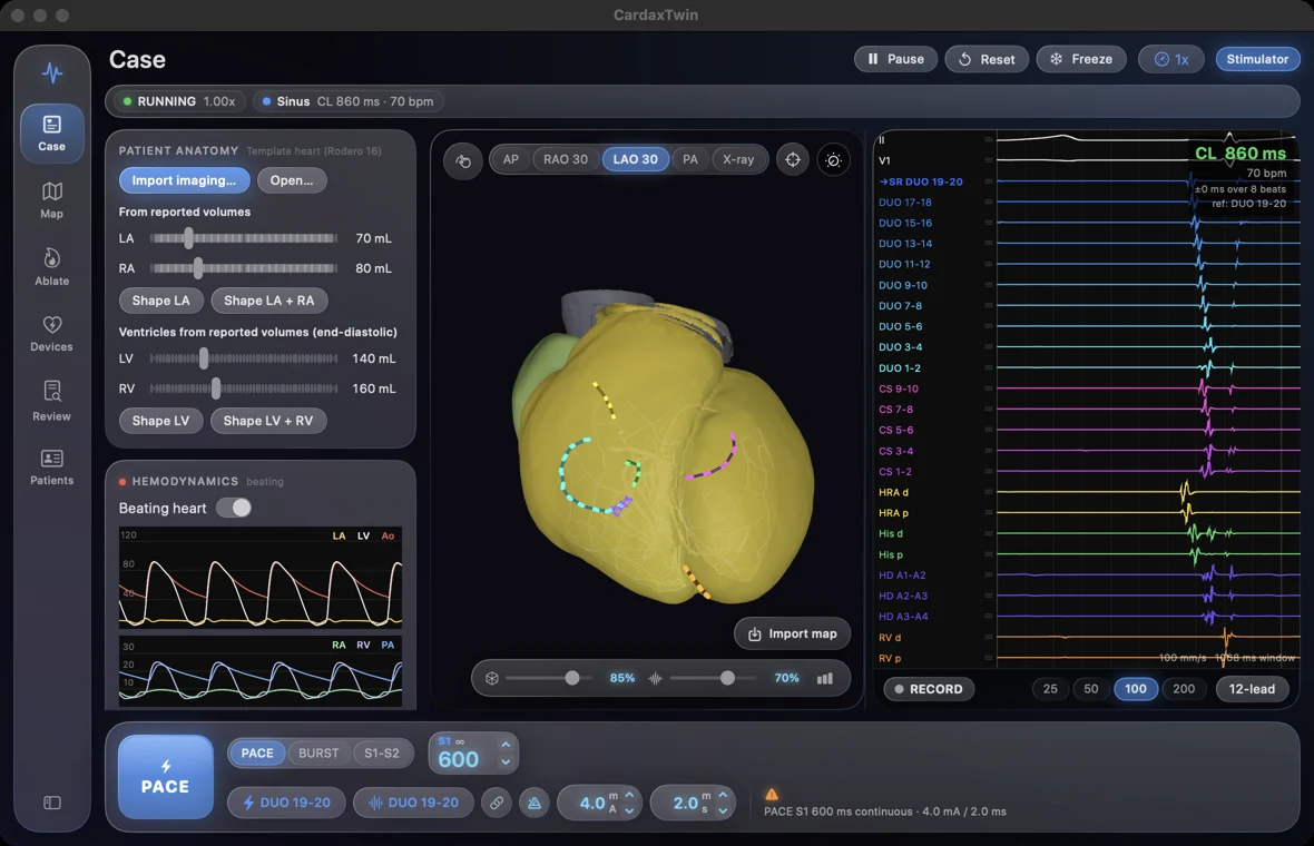
Fit the four-chamber anatomy
Start with the CardaxEP template or use prepared imaging to fit atrial and ventricular anatomy. Chamber measurements provide another starting point when surfaces are unavailable.
- Fit the atria to prepared CT or MRI surfaces or estimate geometry from reported chamber volumes.
- Register the ventricular model against left and right ventricular surfaces and the epicardium.
- Rebuild conduction structures, fiber directions, and ECG lead fields on the fitted anatomy.
Review imported study measurements
Inspect reported values before applying them to a model. The review shows what each selected value can change and reports differences that remain after the fit.
- Review chamber volumes, wall thickness, and other dimensions alongside the model.
- Review hemodynamic inputs such as pressure and cardiac output before fitting the circulation.
- Reopen saved research cases and compare fitted values with the original measurements.
Model scar and voltage substrate
Explore how the location and depth of modeled scar affect activation. Imaging and voltage maps provide different kinds of information and can inform different parts of the model.
- Use supported late gadolinium enhancement MRI inputs to place modeled dense scar and border zone.
- Explore CT wall thinning as a research proxy for scar where supported.
- Use imported electroanatomic voltage data to inform the endocardial substrate and inspect the resulting activation.
Connect activation, motion, and circulation
Follow a simulated beat from electrical activation to wall motion and hemodynamics. The circulation can be fitted after the chamber geometry is established.
- Visualize electrically driven contraction of the beating heart.
- Inspect simulated pressures, chamber volumes, and pressure-volume loops.
- Compare the model’s pressures, volumes, or output with the source measurements used in the fit.
Run a virtual EP study
Use the familiar CardaxEP laboratory on the fitted heart. Change one part of the model and watch how the rhythm or recording responds.
- Position virtual catheters and inspect intracardiac electrograms alongside the simulated 12-lead ECG.
- Use programmed stimulation, activation maps, and simulated ablation to explore possible mechanisms.
- Compare template anatomy with a fitted model to see where geometry and substrate change the result.
Explore virtual cohorts
Run research experiments across synthetic cases. The virtual cohort tools let you vary the model systematically and keep simulated records distinct from clinical data.
- Vary chamber size, scar distribution, and circulation assumptions across modeled cases.
- Compare activation timing, ECG features, and modeled responses to stimulation.
- Rerun a synthetic case with one assumption changed to see which outputs are sensitive to it.
Interpret results in context
CardaxEP Twin is a research environment. A fitted model is an approximation, and a simulated arrhythmia is a result to investigate.
- Keep observed measurements, fitted values, and modeling assumptions distinct.
- Use fit residuals to identify measurements the model does not reproduce.
- Use the output for simulation research; it is not validated for diagnosis, treatment planning, or prediction of individual clinical outcomes.
Research alpha
Beating heart in sinus rhythm
A rotating view of the synthetic four-chamber model shows the heart beating in simulated sinus rhythm.
Open videoScar-related ventricular tachycardia
Follow activation bars around the ventricle in a simulated scar-related VT example.
Open videoTypical atrial flutter
Colored activation bars make the repeating atrial wavefront visible on the beating model.
Open videoAtrial fibrillation
Watch shifting activation and wavebreak in the atria of a synthetic model.
Open video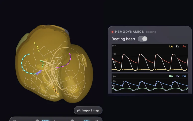
Activation alongside pressure traces
The simulation shows conduction in the template heart next to modeled atrial, ventricular, and arterial pressures.
Open full image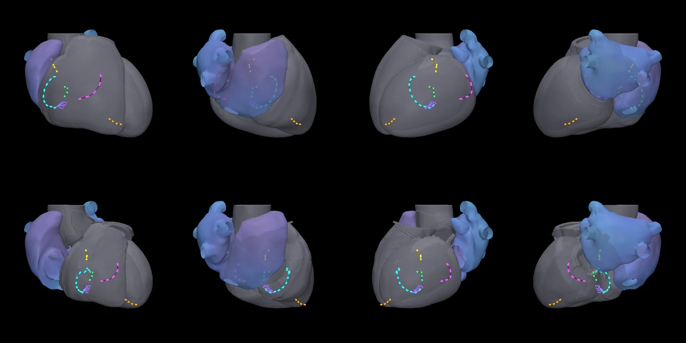
Wall motion and catheter position
Compare views across phases of the simulated beat to see how the atria, vessels, and virtual catheter positions move with the heart.
Open full image