
ICE
Steer a virtual intracardiac echo catheter and see how its position changes the ultrasound view.
macOS workstation and CardaxEP desktop integration
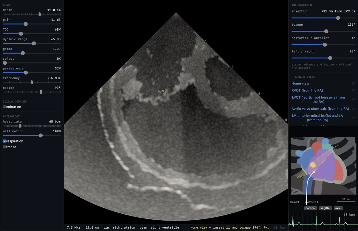
Catheter navigation and orientation
Relate the angle of the ultrasound beam to the catheter position and surrounding anatomy.
- Advance and withdraw the catheter, rotate its shaft, and adjust deflection along two axes.
- Start in the home view or follow the simulated femoral, iliac, and caval route into the right atrium.
- Follow the path through the tricuspid valve, right ventricle, outflow tract, and pulmonary artery.
- Use the anatomy navigator and tip and beam readouts to understand what sits inside and outside the imaging plane.
Standard views and landmarks
Use presets to study the landmarks, then change the catheter position to see which structures come into plane.
- Compare the home view, caval and hepatic views, and the interatrial septum and fossa ovalis.
- Explore short- and long-axis views of the aortic valve, the left ventricular outflow tract, and the mitral valve.
- Locate the left atrial appendage, pulmonary veins, cavotricuspid isthmus, and right ventricular outflow tract.
- Compare right ventricular and pulmonary artery views of left-sided structures.
Ultrasound image controls
The simulator forms the ultrasound sector from an acoustic tissue model. Catheter motion changes the anatomy that the beam crosses.
- Set imaging depth, gain, time-gain compensation, dynamic range, frequency, and sector width.
- Tune the grayscale display with reject, persistence, gamma, and grayscale presets.
- Freeze the image and step through the cardiac phase to compare wall and valve motion.
- Explore how simulated tissue boundaries, instruments, and imaging artifacts affect the sector.
Color Doppler
See how the same simulated flow can appear differently when viewed from another position or with another velocity scale.
- Inspect modeled flow through valves, outflow tracts, and veins during different phases of the heartbeat.
- Move, resize, or change the depth of the color box and adjust the gain and wall filter.
- Vary the Nyquist limit to see aliasing and compare flow toward and away from the transducer.
Transseptal simulation
Rehearse the visual sequence of aligning the sheath, observing septal tenting, and following the simulated crossing.
- Insert and steer the virtual sheath and compare contact at the fossa with contact at the muscular rim.
- Use ICE to bring the needle into plane and compare the visible tip with an out-of-plane position.
- Observe the sheath after a simulated crossing and see how the guidewire curls when it has room in the left atrium.
Wall contact and rhythm-linked motion
Use feedback from the model to connect catheter movements, cardiac phase, and the appearance of the image.
- Distinguish a free tip from one sliding along or pressing against a wall.
- Explore modeled wall perforation and pericardial effusion in the simulator.
- Change standalone heart rate, wall motion, and respiration or observe a linked CardaxEP simulation rhythm.
Simulation and education
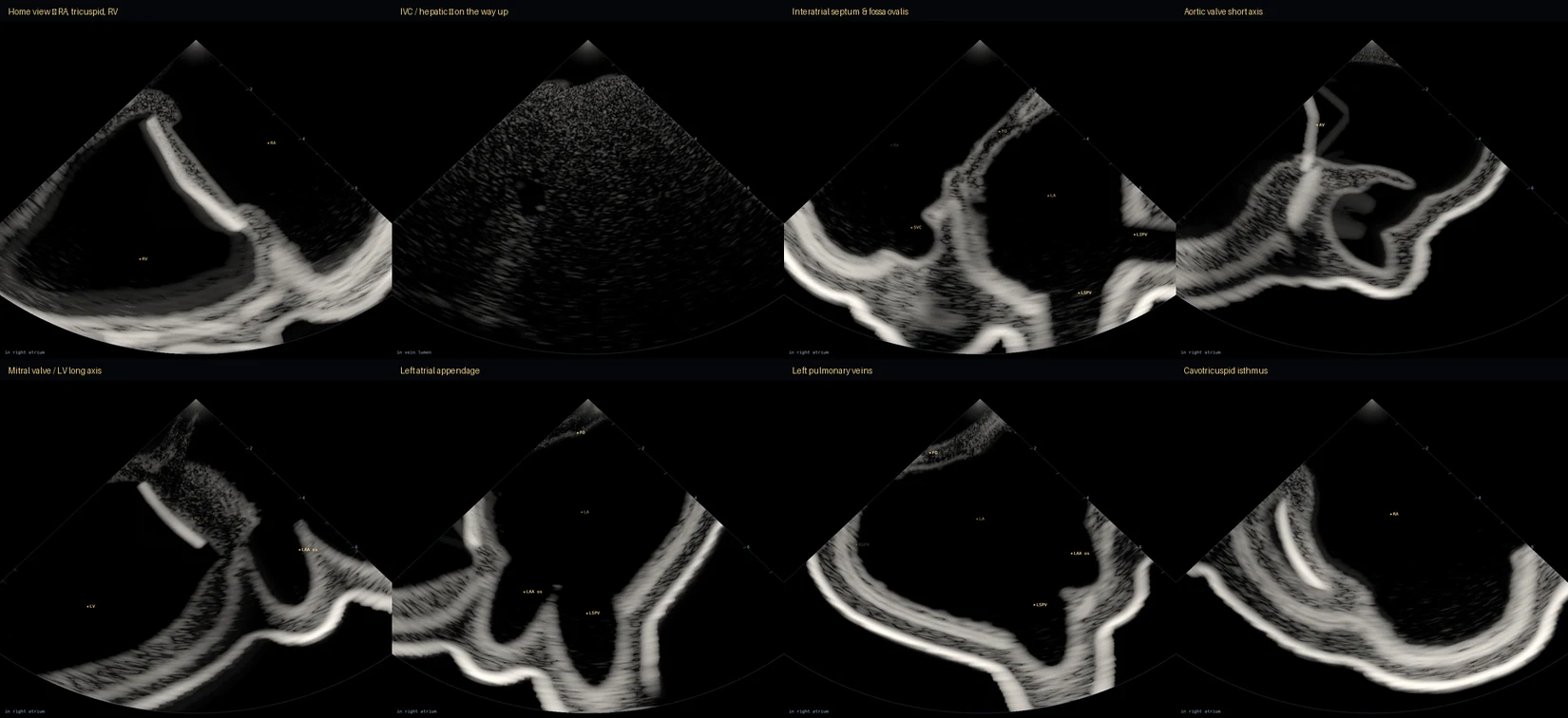
Standard view atlas
Eight simulator views, including the home view, fossa ovalis, aortic valve, left atrial appendage, and pulmonary veins.
Open full image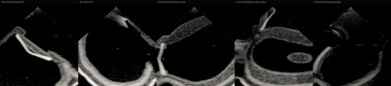
Imaging from the right heart
Five views compare the anatomy visible from the atrium, ventricle, and pulmonary artery.
Open full image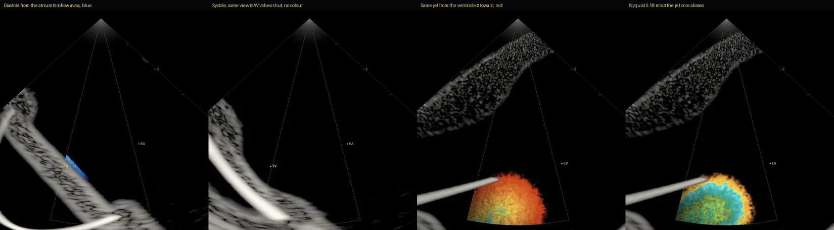
Color Doppler and aliasing
Compare different viewing angles and the effect of changing the velocity scale on a simulated flow jet.
Open full image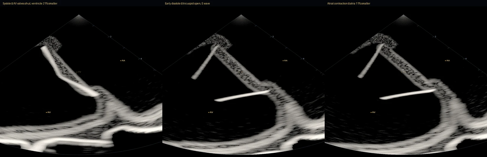
Three phases of the heartbeat
Compare simulated wall and valve motion during systole, early diastole, and atrial contraction.
Open full image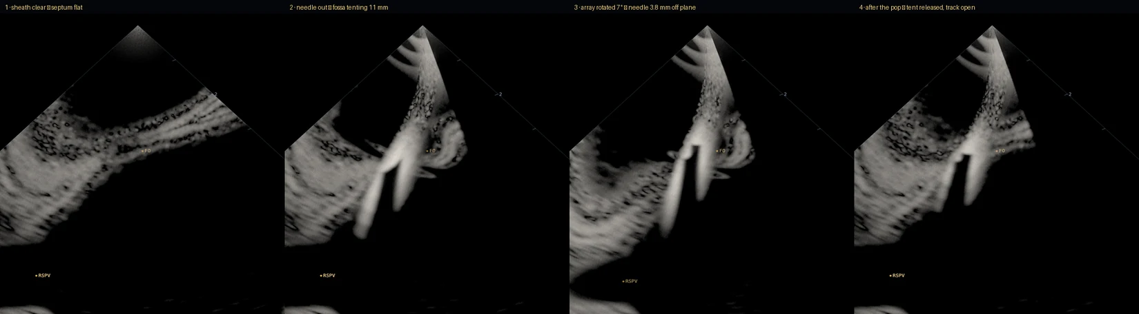
Septal tenting and needle alignment
Follow the simulated sheath from first contact to tenting, then compare how the needle appears in and out of plane.
Open full image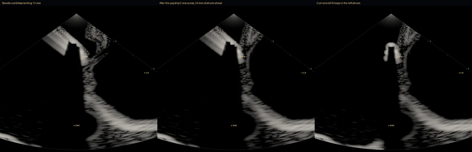
Guidewire confirmation
Three frames show the sheath near the septum and the curled guidewire after a simulated crossing.
Open full imageModified anatomy: Rodero, Strocchi, Marciniak et al., Virtual cohort of adult healthy four-chamber heart meshes from CT images (Zenodo, 2021, doi:10.5281/zenodo.4590294), CC BY 4.0. BodyParts3D, © The Database Center for Life Science licensed under CC Attribution 4.0 International. Heart anatomy dataset · BodyParts3D attribution · CC BY 4.0 license