
CardaxLab
Compare radiofrequency and pulsed-field ablation in a virtual device research bench.
macOS
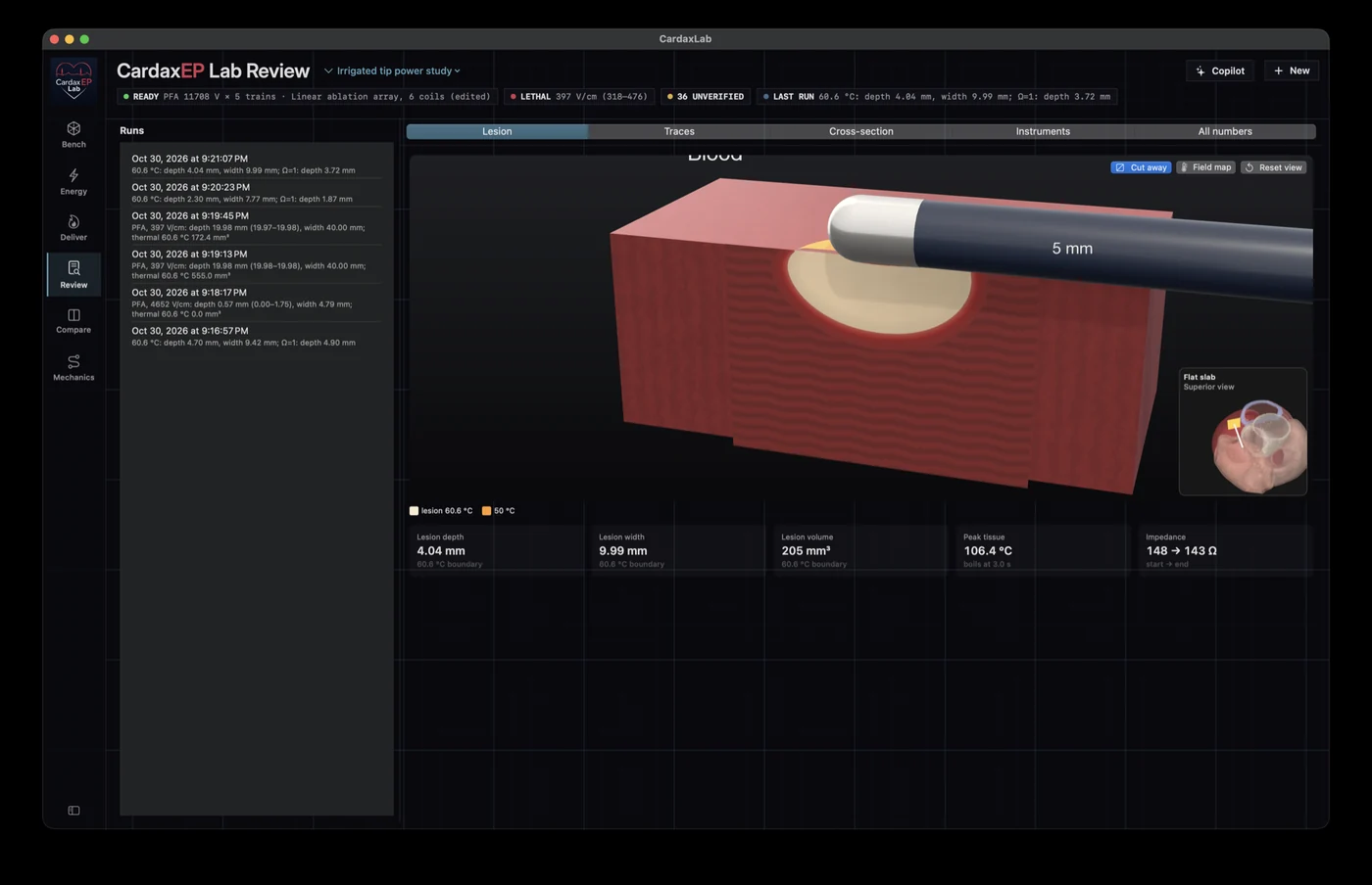
Build the virtual bench
Define the tissue, catheter, and energy settings for an engineering question. Start with a simple tissue slab or explore tissue blocks that represent specific anatomic scenarios.
- Set the tissue properties, wall thickness, surrounding fluid, and cooling assumptions.
- Choose a device form and adjust catheter orientation and contact.
- Explore modeled septal, left ventricular summit, and posterior left atrial scenarios.
Radiofrequency ablation
Run a rotationally symmetric bench or a three-dimensional simulation. Follow the modeled current, heat transfer, and thermal injury through delivery and cooling.
- Compare irrigated and nonirrigated electrode setups.
- Explore power, voltage, and temperature control in supported generator models.
- Inspect temperature contours, thermal damage boundaries, and lesion cross sections.
Pulsed-field ablation
Explore how electrode arrangement and pulse delivery change the modeled electric field. The bench can show predicted exposure and heating separately.
- Configure pulse amplitude and train structure for supported device models.
- Compare field contours and modeled electroporation thresholds.
- Inspect the thermal contribution of pulse delivery alongside the electric field.
Catheter geometry and contact
Explore the mechanical and electrical consequences of changing the device or its position before committing to a physical bench experiment.
- Explore supported tip, loop, and grid electrode forms.
- Vary contact, insertion, and angle, and inspect the modeled tissue interface.
- Compare electrode spacing and the effect of orientation on the field.
Virtual instruments and run review
Watch traces during a run, then inspect the result at multiple scales. Review a cross section to see where a threshold falls in the tissue.
- Inspect temperature maps and simulated temperature probes at different depths.
- Review simulated power, energy, and impedance traces where available.
- Use cross sections and 3D views together to inspect heating, field exposure, and modeled lesion geometry.
Compare experiments side by side
Use the workbench to evaluate a change in setup against a previous run. Compare the parameter differences and results in the same view.
- Compare lesion and energy measurements alongside the settings that produced them.
- Overlay traces and display cross sections on a shared color scale.
- Use CPU and supported Metal GPU solvers for applicable three-dimensional experiments.
Track parameters and their sources
Keep each study’s settings and assumptions attached to its runs. Review proposed parameters before they are used.
- Record a value, its unit, and where it came from.
- Review or reject suggestions from the optional local parameter assistant.
- Keep defaulted and unverified values visible when interpreting a run.
Validation and scope
The workbench includes a validation workspace for reviewing benchmark questions, predicted ranges, and comparisons with measurements. Results depend on the modeled conditions.
- Inspect benchmark cases, comparison rules, and recorded verdicts.
- Use the RF and pulsed-field benches to explore relative effects and plan research experiments.
- Mapping, pacing, and defibrillation are broader research directions. The ablation results are not validated for clinical decisions.
Engineering research
Inside the CardaxLab bench
A silent tour of three views: an RF lesion model, a virtual thermal camera with temperature traces, and two pulsed-field configurations.
Open video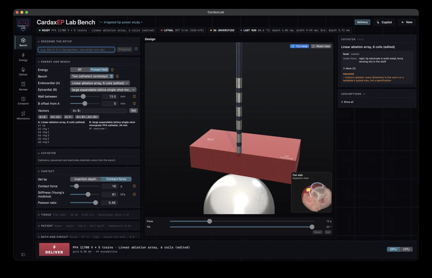
Bench setup
The setup places the device against a tissue block while the inspector exposes the settings and assumptions for the run.
Open full image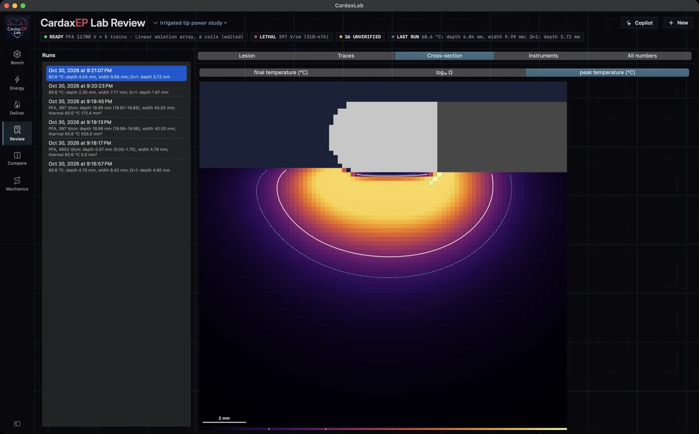
RF lesion cross section
The cut plane reveals temperature and damage contours beneath the virtual electrode.
Open full image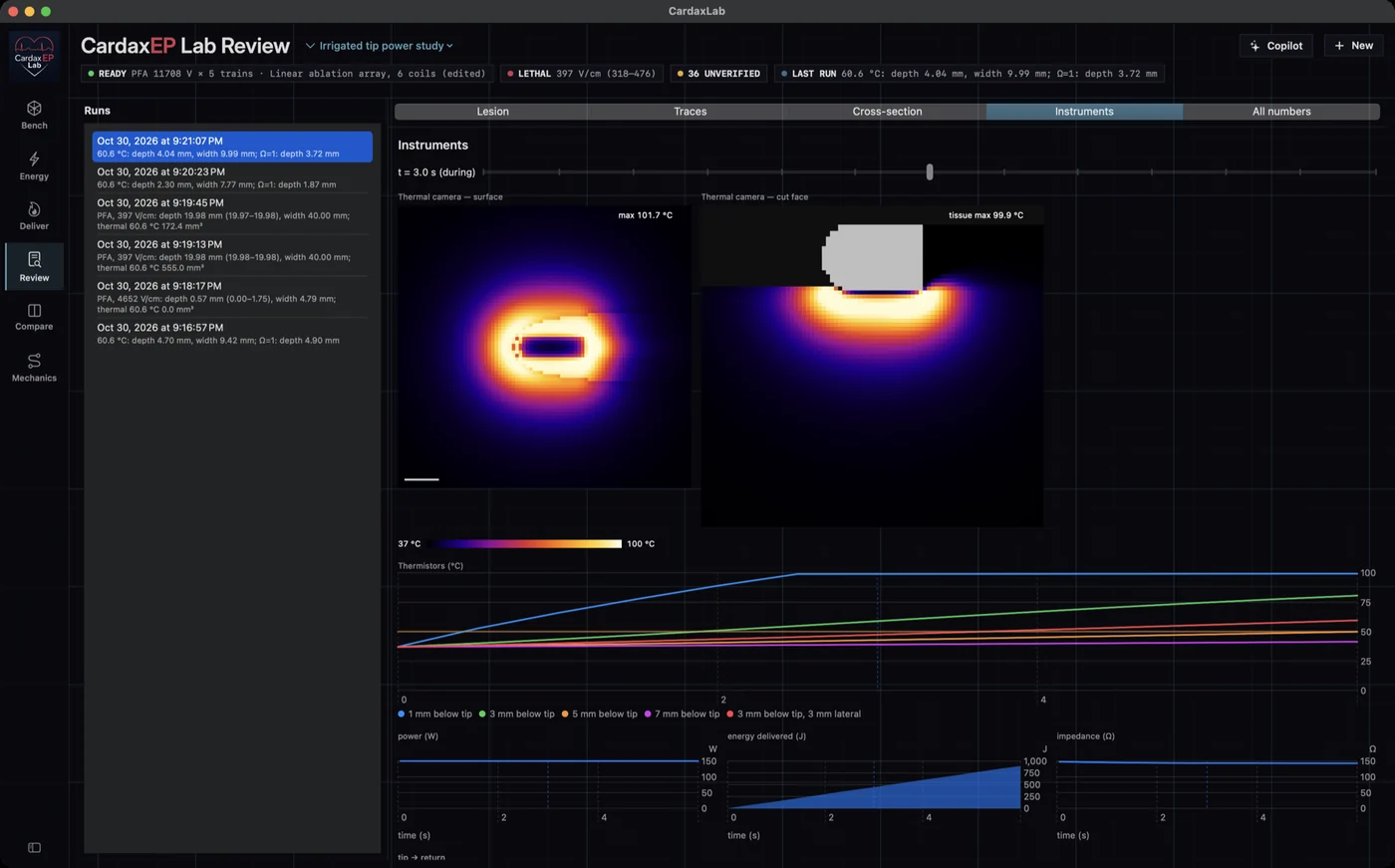
Temperature maps and traces
Surface and cross-sectional thermal views sit above the simulated instrument traces.
Open full image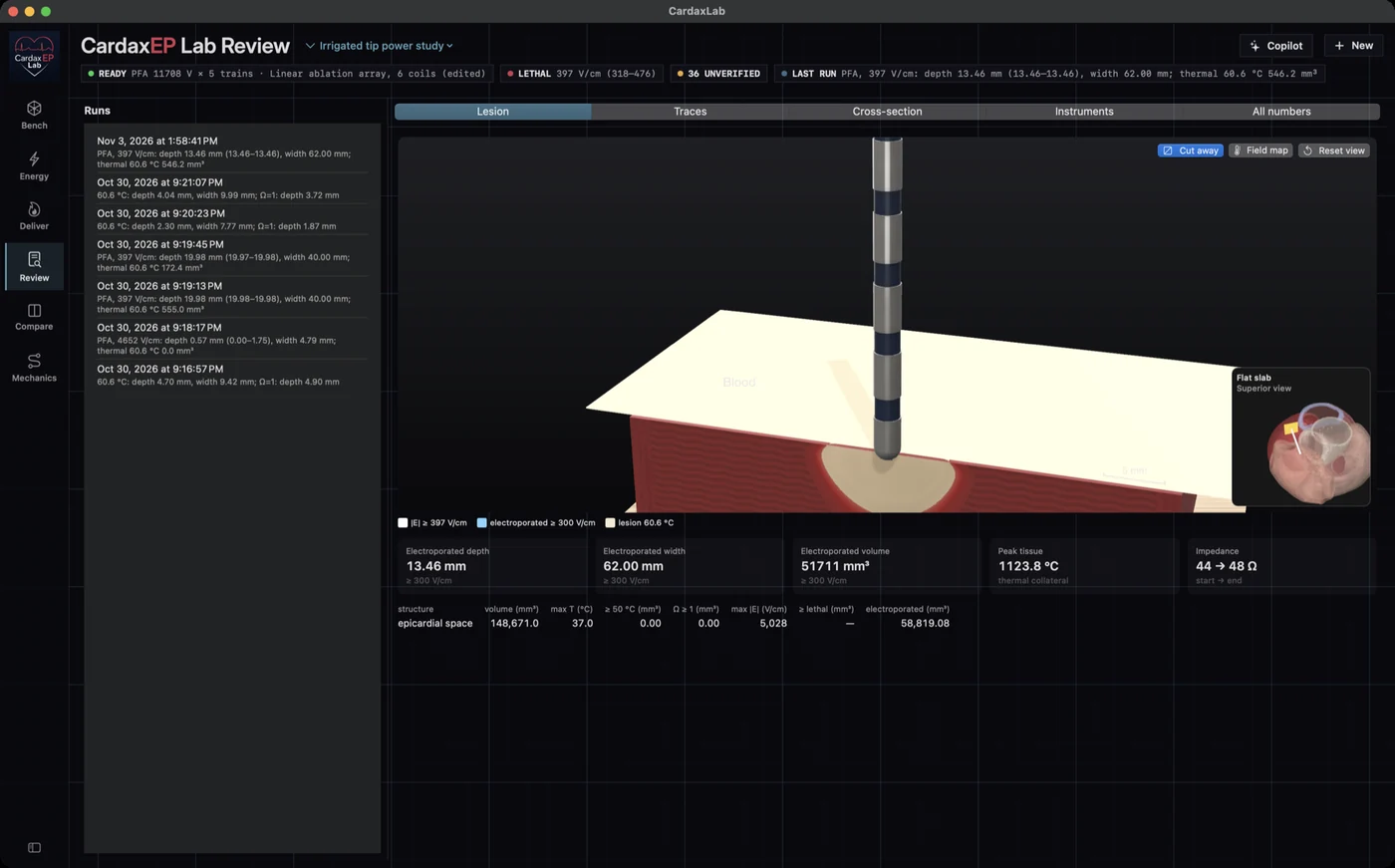
Pulsed-field run review
The workbench shows an electrode array, tissue block, and modeled exposure for a pulsed-field experiment.
Open full image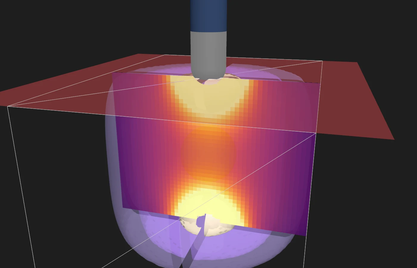
Pulsed-field septal bench
A synthetic tissue cross section shows the modeled field between electrodes on opposite sides of the septum.
Open full image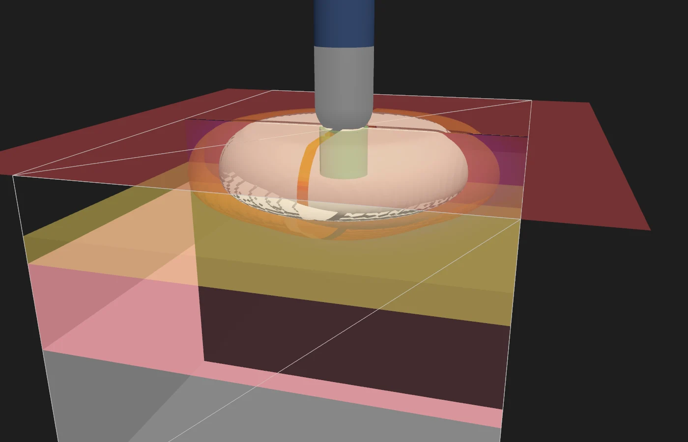
Posterior left atrial RF scenario
The synthetic bench places an RF application near a modeled neighboring structure to visualize the thermal region.
Open full image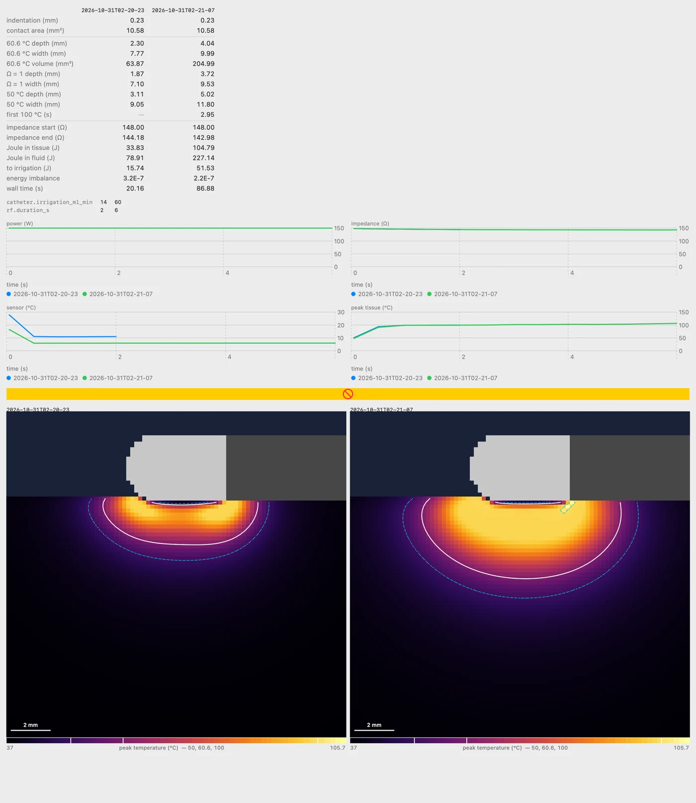
Side-by-side run comparison
Parameter values, traces, and RF cross sections share one view so differences between two runs are easier to inspect.
Open full image